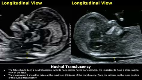
nuchal fold thickness normal measurement|nuchal fold measurement chart : store The nuchal fold is a normal fold of skin at the back of a baby’s neck. This can be measured between 15 to 20 weeks in pregnancy as part of a routine prenatal ultrasound. The nuchal fold .
WEBJesus para crianças - Momento Espírita Filmes @canalfep . LIVRO MOMENTO ESPÍRITA v. 10 . LIVRO MOMENTO ESPÍRITA v. 5 . Momento Espírita estréia na TV BAND . MOMENTO ESPÍRITA NO .
{plog:ftitle_list}
Felix Gaming's highly-playable, fun slots are only found at a handful of casinos. Their games come with standard welcome bonuses that are often topped with free spins. Here are the best online casinos to feature Felix Gaming titles.
The nuchal fold is a normal fold of skin seen at the back of the fetal neck during the second trimester of pregnancy. Increased thickness of the nuchal fold is a soft marker associated with multiple fetal anomalies, and is measured on a routine second trimester .What is the normal range of nuchal fold thickness at different stages of pregnancy? The normal range of nuchal fold thickness varies as pregnancy progresses. Here’s a quick guide: Comparison of nuchal skin fold thickness (NFT) in a normal 20-week fetus on the standard horizontal transcerebellar and 30° occiput images. (a) The standard horizontal . Second trimester thickened nuchal fold has a high specificity for aneuploidy. ACOG and SMFM define an abnormal nuchal fold as ≥ 6mm between 15 and 20 weeks of gestation. It is the most powerful second .
Increased measurement of the nuchal fold (≥ 6 mm from 14 weeks to 22 weeks of gestational age) is considered a soft marker for chromosomal aneuplodies, as well as for structural defects in the fetus, most commonly cardiac defects.The nuchal fold is a normal fold of skin at the back of a baby’s neck. This can be measured between 15 to 20 weeks in pregnancy as part of a routine prenatal ultrasound. The nuchal fold .
Normal thickness: All fetuses develop a measurable nuchal translucency at some point in the first trimester. Thickness of the translucency varies with gestational age: Peak thickness at 12-13 weeks (in 75% of fetuses). At 12-13 .Nuchal fold can be spuriously thickened by angling caudally (intersecting the inferior level of the cerebellum and occiput). This nuchal skin fold increases with advancing gestational age and . Current guidelines from the American College of Obstetricians and Gynecologists (ACOG) and the Society for Maternal-Fetal Medicine (SMFM) state that screening (serum .
Our nuchal translucency calculator finds the percentile of the nuchal fold thickness and compares it with the nuchal translucency measurement chart. This calculation allows us to estimate the risk of the .Prenatal ultrasound: Increased nuchal fold (2nd trimester)
Nuchal Translucency Normal Range Chart. When the nuchal scan is done, the doctor will share the results with you. At that time, it is important to understand what a normal measurement is. For a baby that is between 45 . Nuchal translucency (NT) is a measure of a thickness of a fold located on the fetuses' neck. This fold's greater thickness is connected to the greater prevalence of genetic disorders, fetal death, and its major .What is Nuchal Translucency (NT)? NT is the name given to the black area seen by ultrasound at the back of the fetal head/neck between 11 - 14 weeks of gestation. The NT represents a normal accumulation of fluid, but, if too thick (usually above 3-3,5mm), it is a sign that something may not be going well with the development of your baby.
Normal NT measurements vary depending on how far along you are in your pregnancy. In general, most doctors consider a normal NT measurement at 12 weeks to be under 3 millimeters. . Then he or she will locate the nuchal fold and measure its thickness on the screen. Those measurements, plus your age and baby's gestational age, will be entered .The nuchal translucency test measures the nuchal fold thickness. This is an area of tissue at the back of an unborn baby's neck. . How the Test is Performed. Your health care provider uses abdominal ultrasound or a vaginal ultrasound to measure the nuchal fold. All unborn babies have some fluid at the back of their neck. . A normal amount .This nuchal skin fold increases with advancing gestational age and ranges between 1 and 5 mm in normal fetuses between 14 and 21 weeks gestation. A nuchal skin fold thickness of ³ 6 mm is . , Fassnacht MA et.al. Routine measurement of nuchal thickness in the second trimester. J Matern Fetal Med 1992; 1:82-86 ;
The nuchal translucency test measures the nuchal fold thickness. This is an area of tissue at the back of an unborn baby's neck. . Your health care provider uses abdominal ultrasound or a vaginal ultrasound to measure the nuchal fold. All unborn babies have some fluid at the back of their neck. . there is more fluid than normal. This makes .The nuchal translucency test measures the nuchal fold thickness. This is an area of tissue at the back of an unborn baby's neck. . Your health care provider uses abdominal ultrasound or a vaginal ultrasound to measure the nuchal fold. All unborn babies have some fluid at the back of their neck. . A normal amount of fluid in the back of the .Nuchal Fold Thickness -The Nuchal fold is a regular fold of skin found at the back of the fetal neck during the second trimester of pregnancy. Increased nuchal fold thickness is a soft indicator associated with a variety of fetal abnormalities and is measured during a regular second-trimester ultrasound. . Even a normal NT measurement . While both measurements are at the level of the fetal head or neck, a nuchal fold thickness, which is only performed in the second trimester, should not be confused with a first trimester nuchal translucency (NT) measurement. If an enlarged second trimester nuchal fold measurement is obtained, next steps should include. Detailed anatomic study
An abnormal measurement is when the skin fold measure is larger than the normal range of up to 2 mm at 11 weeks or 2.8 mm at 13 weeks 6 days. Your doctor will consider the measurements along with .
A measurement of 2 mm to 10 mm is normal in the second and third trimesters. The nuchal fold is a measurement taken from the outer skin line to the outer bone in the midline. Less than 6 mm is considered normal up to 22 weeks. . A nuchal fold thickness of >6 mm is considered abnormal and is seen in 80% of newborns with Down syndrome. Cystic .What is a normal nuchal translucency measurement? An NT of less than 3.5mm is considered normal when your baby measures between 45mm and 84mm from crown to rump (PHE 2018). The NT usually grows in proportion with your baby (Nicolaides 2011). The images below give an idea of what different levels of NT look like.
Normal thickness: All fetuses develop a measurable nuchal translucency at some point in the first trimester. Thickness of the translucency varies with gestational age: Peak thickness at 12-13 weeks (in 75% of fetuses). At 12-13 .To assess the association of adverse pregnancy outcomes in fetuses with increased nuchal folds in the setting of normal genetic testing. Increased measurement of the nuchal fold (≥ 6 mm from 14 weeks to 22 weeks of .Objective: To establish normal values of fetal nuchal fold thick-ness at 14-16 weeks of gestation by transvaginal sonography. Methods: Transvaginal sonography was used to measure nuchal fold thickness in 182 normal pregnancies at 14-16 weeks of gestation. Nuchal fold thickness was measured as the distance from the outer skull bone to the outer skin surface in the .
when to measure nuchal fold
Around the 12th week of gestation, a doctor can measure the fetus’s nuchal fold and assess whether it’s larger than expected. Nuchal fold measurement. The nuchal fold measurement, also known as the nuchal translucency test, is a non-invasive test that’s performed between the 11th and 14th week of pregnancy. As we’ve mentioned, its . I just got back from my 20 week scan. It looks like baby is measuring big, but everything looked normal except for a nuchal fold measurement of 7mm. We originally opted out of genetic testing but after that measurement decided to go forward.The cut-off for the nuchal translucency measurement is 3.5 mm. If your measurement is less than 3.5 mm, this is considered "normal". If your measurement is 3.5 mm or more, this is considered "increased". . If you were told your NT measurement is "normal" and your prenatal genetic screening results are "screen negative" or "low risk", it means .
Comparison of nuchal skin fold thickness (NFT) in a normal 20-week fetus on the standard horizontal transcerebellar and 30° occiput images. (a) The standard horizontal transcerebellar image of a normal 20-week fetus gives a NFT measurement of 6.5 mm.
A nuchal translucency (NT), also called nuchal fold or nuchal thickness, is a measurable area at the back of the fetal neck. It is examined using ultrasound as part of combined screening for Down, Edwards and Patau syndromes from 11 to 13 weeks and six days of pregnancy.. To measure the NT, the fetal crown-rump length should be between 45 millimetres and 84 . A thickened nuchal fold is defined as ≥6 mm between 15 and 20 weeks of gestation. 99 Thickened nuchal fold was one of the first identified ultrasound markers of trisomy 21 and is one of the most specific markers to date. 99, 100 After the initial report, several prospective series of consecutive amniocenteses confirmed the association between . The result of semen analysis indicated low motility (35%) and high abnormal morphology (88%). During the routine first trimester screening at 13 weeks of gestation, NT was measured at 3 mm. The normal range of NT for this age is 1.6-2.4 mm. Nuchal skin fold (NF) measurements and prenatal follow-up ultrasound findings were normal. Comparison of nuchal skin fold thickness (NFT) in a normal 20-week fetus on the standard horizontal transcerebellar and 30° occiput images. (a) The standard horizontal transcerebellar image of a normal 20-week fetus gives a NFT measurement of 6.5 mm.
To compare the measurements of fetal nuchal fold (NF) thickness by two-dimensional (2D) and three-dimensional (3D) ultrasonography using the three-dimensional extended imaging (3DXI). Methods. A cross-sectional study was performed with 60 healthy pregnant women with a gestational age between 16 and 20 weeks and 6 days.While a nasal bone may be absent in some fetuses with a chromosomal abnormality, most with this finding are normal. Combining your age-related risk with the nuchal translucency measurement, nasal bone data and bloodwork provides one risk result for Down syndrome and a separate risk result for trisomy 13 or trisomy 18.

tramex cmexpert ii digital concrete moisture meter
tramex compact moisture meter
WEB1000 Blocks. Courage Saw Game. Flow Free Online. Classic Tetris. Minesweeper Game. Butterfly Kyodai. Vampire Skills. Mahjong Alchemy. Kris Mahjong. Snail Bob. Jewel .
nuchal fold thickness normal measurement|nuchal fold measurement chart