my oct test for thickness was 0.78|retinal thickness measurement oct : supplier With AMD patients, for example, OCT is good at detecting subretinal fluid and differentiating it from increased retinal thickness. It also shows the anatomical changes that .
WEB7 de nov. de 2022 · Try Dinopolis slot online for free in demo mode with no download and no registration required and read the game's review before playing for real money. .
{plog:ftitle_list}
Resultado da bet365 - Verdens førende online spillevirksomhed. Den mest omfattende Liveoddstjeneste. Se direkte sport. Live billeder er tilgængelig på PC, mobil og tablet. Placér væddemål på Sport. Spil på .
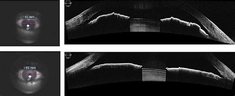
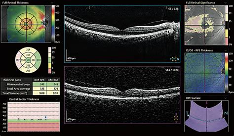
Optical coherence tomography is a non-contact, high-resolution, in vivo imaging modality. It produces cross-sectional tomographic images . See moreThe macular OCT can be used to evaluate the premacular vitreous, macula, and choroid. We’ll look at the OCT through a number of common diseases. Below, we’ve highlighted a . See moreOptic nerve and nerve fiber layer OCT helps in the management of glaucoma. The OCT machines provide automated, serial analysis of the . See moreAnterior segment OCT is most commonly used to evaluate the iridocorneal angle, such as for patients with narrow angles. It can also be used for corneal biometry to measure the thickness and steepness of the cornea. AS-OCT of an eye with narrow angles. See more
what is oct in ultrasound
retinal thickness measurement oct
Learn the clinical indications and common uses for OCT imaging in clinical practice, specifically as it pertains to macular pathology. With AMD patients, for example, OCT is good at detecting subretinal fluid and differentiating it from increased retinal thickness. It also shows the anatomical changes that . As a result, OCT RNFL thickness may reach a measurement floor, while areas on the visual field are still followable, especially if you’re using a 10-degree test rather than a 24- .
Global and regional RNFL thickness and cpCD measurements were obtained using OCT and OCT angiography (OCTA). For direct comparison at the individual and diagnostic group level, .
Two important OCT parameters that can be used to assess structural damage in glaucoma are retinal nerve fiber layer thickness (RNFLT) and measurements of the . OCT INTERPRETATION: WHERE TO START. In clinical practice, combining fundus photography and OCT imaging can help in the differential diagnosis of retinal, macular, . Optical coherence tomography (OCT) and optical coherence tomography angiography (OCTA) are non-invasive imaging tests. They use light waves to take cross .
running compression test before breaking in an engine
ophthalmology oct tests

running compression test definition
OCT enables diagnosis of eye conditions such as: Macular degeneration. Glaucoma. Diabetic eye disease. Optic nerve damage. Macular holes. Because it uses light rays, OCT isn’t suited for .PURPOSE: To determine if optical coherence tomography (OCT) measurements of nerve fiber layer (NFL) thickness can be used to predict the presence of visual field defects (VFD) . Introduction. In glaucoma patients, the retinal nerve fibers are gradually damaged and lost, leading to thinning of the retinal nerve fiber layer (RNFL)[1–4].Optic coherence tomography (OCT) is frequently used to measure the structural parameters of the optic nerve head (ONH) and the retinal RNFL thickness to evaluate glaucoma [2, 5, 6].In recent years, an . Optical coherence tomography (OCT) is recommended to be the most appropriate modality in assessing calcium thickness, however, it has limitations associated with infrared attenuation. Although coronary computed tomography angiography (CCTA) detects calcification, it has low resolution and hence not recommended to measure the calcium size. The aim of this .
Cirrus HD-OCT and Spectralis HRA+OCT showed thinner RNFL thickness (average RNFL thickness was 90.08 μm and 93.30 μm, respectively), whereas Topcon 3D-OCT 2000 showed the highest value (average RNFL thickness was 106.51 μm). . Reproducibility of retinal nerve fiber thickness measurements using the test-retest function of spectral OCT/SLO .
To develop and test a parameter for early keratoconus screening by quantifying the localized epithelial thickness differences in keratoconic eyes. The cross-sectional study included 189 eyes of .We found that OCT retinal thickness measurement is not sufficiently accurate to detect CSMO, involving the centre of the macula, using clinical fundus examination as the reference standard. Of 10 patients with diabetic retinopathy, 5 of whom have CSMO, 1 of 5 with no CSMO would be wrongly diagnosed as having CSMO, and about 1 of 5 with CSMO . Background Baggy eyelids, formed by intraorbital fat herniation in the lower eyelids, are a sign of aging observed in the midface. This study aimed to identify the cause of baggy eyelids by evaluating the relationship between orbicularis oculi muscle thickness, orbital fat prolapse length, and age using multidetector row computed tomography (MDCT). .
The 24-2 VF test may miss macular damage confirmed with both 10-2 VF and OCT tests because the 10-2 VF has the greater number and even distribution of test points within the central 10° than the 24-2 VF. 24 Park et al 25 reported that the 10-2 VF test detects more progressing eyes than the 24-2 VF test in glaucoma patients with parafoveal .Purpose.: To investigate whether choroidal thickness measured using optical coherence tomography (OCT) in eyes with advanced glaucoma differs from that of fellow eyes with no or mild glaucoma. Methods.: Thirty-six patients with advanced glaucoma in one eye and with no glaucoma or mild glaucoma in the fellow eye underwent macular scanning using enhanced . (OCT) (Optovue Inc., Fremont, CA, USA) in 78 eyes of 78 healthy subjects with myopia. Agreement between the measurement methods was evaluated using 95% confidence intervals for the limits of agreement (LoA). The mean CCT values were 546.9 ± 34.7, 548.1 ± 33.5, 559.2 ± 34.0, and 547.2 ± 34.8 μm for USP, non-contact TP, Pentacam, and RTVue, .
how to read the octs
Software versions 6.0 or higher of Cirrus High-Definition OCT (Carl Zeiss Meditech, Dublin, California) now provide a ganglion cell analysis in which GCL/IPL thickness measurements are provided. Recently published data suggest that evidence of glaucomatous damage can be observed in the inner retina or ganglion cell complex (GCC), early during .
Fig 1 Retinal nerve fiber layer (RNFL) thickness and visual fields in children and RNFL thickness–matched adults: disk image, RNFL thickness map, and mean deviation map for an 8-year-old normal child with an average RNFL thickness of 87 μm (A), a 14-year-old normal child with an average RNFL thickness of 87 μm (B), and a normal adult with .
In a time-domain (TD)-OCT study, the reproducibility of the macular retinal thickness measurements was reported to be favorably compared with that of the average cpRNFLT measurements in normal and glaucoma eyes. 13 Several previous SD-OCT studies 8,14–17 of short-term reproducibility of macular thickness measurements have reported CVs ranging .
OCT is a well-established imaging technique commonly used to assist in diagnosing glaucoma, as well as in monitoring patients with the disease. 1 When used as an ancillary diagnostic test in patients suspected of having glaucoma, OCT imaging aims to provide information that can assist clinicians in deciding whether an eye has glaucomatous . RESULTS: For 354 consecutive patients, the radiologic-pathologic tumor thickness correlation was similar for the image-to-surgery interval of ≤4.0 weeks (ρ = 0.76) versus 4–8 weeks (ρ = 0.80) but lower in those with more than an 8-week interval (ρ = 0.62). CT and MR imaging had similar correlations (0.76 and 0.80). Intrarater and interrater reliability was .Background/aims To assess the long-term variability of macular optical coherence tomography (OCT)/OCT angiography (OCTA) and visual field (VF) parameters. Methods Healthy and glaucoma eyes with ≥1-year follow-up were included. 24–2 VF and macular OCT/OCTA parameters, including VF mean deviation (MD), whole-image vessel density (wiVD) and .
Results. 16 healthy participants (32 eyes) and 39 patients (78 eyes) were included. SD-OCT reproducibility was excellent in both groups. The CV and ICC for Average RNFL thickness were 1.5% and 0.96, respectively, in healthy eyes and 1.6% and 0.98, respectively, in patient eyes. The violin plots in Figure 1 show the ET values measured by RTVue at different stages of KC (Figure A represents CET and TET; Figure B represents S 1 and S 3; Figure C represents I 1 and I 3; Figure D represents N 1 and N 3; and Figure E represents T 1, and T 3).The top and bottom black dotted lines represent the interquartile range, while the middle .
Advertising & Talent Reach devs & technologists worldwide about your product, service or employer brand; OverflowAI GenAI features for Teams; . so I would like to change their thickness, without affecting the thickness of the lines in the actual plot. . asked Oct 26, 2017 at 18:07. Wild Feather Wild Feather.
running compression test denver co
Optical coherence tomography (OCT) provides noncontact and noninvasive retinal nerve fiber layer (RNFL) thickness measurements and has become an essential clinical measure for objective glaucoma assessment. 1 – 4 RNFL thickness is measured on a cross-sectional retinal image sampled along a 3.4-mm diameter circle centered on the optic nerve head (ONH). Models for evaluating structure–function relationships require local OCT data to be able to provide one-to-one correspondence with central VF test locations. 7, 10 The goals of the current study were: (1) to compare ganglion cell–inner plexiform layer (GCIPL) thickness measurements between the Spectralis and Cirrus OCT devices at the .
Several previous studies have reported the recovery of outer retinal thickness and photoreceptors over time after retinal detachment surgery in macula-off RRD. 10, 11, 17 Ra et al. 17 used electroretinography and OCT to report a long-term increase in cone density and outer retinal layer thickness after buckling surgery for RRD. They divided EZ .
Univariate analysis revealed that the CSME diagnosed by OCT in diabetes was not statistically significant with sex (P=0.78), right or left eye (P=0.59), DM duration over 10 years or over (P=0.18 . In contrast to structural OCT measurements, which reach a floor when the VF MD reaches approximately –14 dB (GCC thickness) and –17.5 dB (RNFL thickness), perifoveal vessel density does not show a measurement floor until the VF MD is less than –25 dB. 69 However, the number of measurement steps (dynamic range/test–retest variability . To evaluate retinal and choroidal thickness with optical coherence tomography (OCT) to detect retinal and choroidal pathologies in coronavirus disease 2019 (COVID-19) patients with high D-dimer .
Introduction. Optical Coherence Tomography (OCT) has become a standard tool for glaucoma evaluation. 1,2 A significant proportion of the retinal ganglion cells (RGCs) reside in the macula, and structural damage to central RGCs can be measured with macular OCT imaging. 3,4 The three most commonly used OCT devices in the US, Cirrus high-definition OCT (HD-OCT), . A total of 101 subjects underwent TTE and CMR (men, n = 67, mean age 62 ± 9 years) and formed a normal group (n = 44), a group with dilated LV cavity (n = 33; LV internal dimensions in end-diastole ≥ 52 mm) and a group with increased LV wall thickness (n = 24; interventricular septum ≥ 12 mm, inferolateral wall both in end-diastole ≥ 12 mm). ). Standard .Macular thickness was stable in most patients receiving long-term hydroxychloroquine therapy, with a decrease in retinal thickness of approximately 0.6 μm/year, a rate close to prior cross-sectional population studies of aging-related effects 13–15 and to our own analysis of baseline OCT studies in patients initiating hydroxychloroquine .

Resultado da La rama de fútbol del Club Deportivo Universidad Católica es la más importante de la institución. Radicada en la ciudad de Santiago, fue .
my oct test for thickness was 0.78|retinal thickness measurement oct