measurement of small intestine thickness cats|normal cat stomach ultrasound : manufacture The ultrasonographic and histopathological features of benign fibrotic small intestinal strictures have recently been described in eight cats with chronic intestinal obstruction. 70 The ages of the cats ranged from 2–10 . Andressa iniciou na atividade por acaso. Ela saiu da casa dos pais aos 15, após desavenças familiares (já resolvidas, segundo ela), e foi morar com a tia. No terreno havia um lava-jato de automóveis, e seu primeiro emprego acabou sendo lá, lavando carros. Certo dia, um caminhoneiro encostou o veículo na . Ver mais
{plog:ftitle_list}
WEB23 de dez. de 2023 · Torinha s Porno Grátis - confira essa entre outras cenas Torinha s exclusivas arada e bem safada essa gata deu no couro com esse sacana que te deu uma penetrada das boas na xoxota deliciosa e bem gostosa. . Bem vindo, Se você está em busca das melhores fotos e videos do onlyfans, privacy,fansly temos os melhores .
testing a steel bar with universal testing machine
The normal sonographic thickness of the individual layers (ie, mucosa, submucosa, muscularis and subserosa-serosa) of the intestinal wall was evaluated in 20 clinically healthy cats. The mean thickness of the wall was 2.20, 2.22, 3.00 and 2.04 mm for duodenum, jejunum, ileum (fold) . B-mode ultrasonography is likely the most widely used modality for imaging the gastrointestinal (GI) tract in cats, providing information on intestinal wall thickness and the presence or absence of intestinal dilation and .
The ultrasonographic and histopathological features of benign fibrotic small intestinal strictures have recently been described in eight cats with chronic intestinal obstruction. 70 The ages of the cats ranged from 2–10 .In normal dogs and cats, the small intestines are relatively uniform in distribution. Depending on the segment of small intestine, some layers may be thicker than others. This can be used to identify the different segments of .Note: Normal ultrasonographic measurements of the individual layers of the canine11 and feline7,12 gastrointestinal tract have been described in recent literature. BOX 1 Criteria for .Lymphoma is the most common intestinal neoplasm in the cat. It appears as a focal mass, multiple masses, or diffuse infiltrative neoplasia; characterized by thickening and/or loss of the .
testing in universal testing machine civil
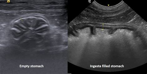
It usually measures between 3 and 5 mm between rugae. A wall thickness greater than 7 mm is considered abnormal in a noncontracted status. The mean number of gastric peristaltic .An ultrasound image showing 2 sections of small intestine. Two callipers (crosses with linked dotted line) have been placed to measure intestinal wall thickness, from the inner edge of the mucosa outwards to the outer edge of .B-mode ultrasonography is likely the most widely used modality for imaging the gastrointestinal (GI) tract in cats, providing information on intestinal wall thickness and the presence or.Note the overall thickening of the small intestine at 3.2 mm (normal is < 2.5 mm) and the thickness of the muscularis layer in particular at 1.5 mm. Chronic enteropathies are a common clinical problem in cats, and in most cases are .
maximum thickness of 4 mm on the transverse view.13 In normal dogs and cats, the small intestines are relatively uniform in distribution. Depending on the segment of small intestine, some layers may be thicker than others. This can be used to identify the different segments of intestines. For example, in the dog, the mucosal layer of theserosa) of the intestinal wall was evaluated in 20 clinically healthy cats. The mean thickness of the wall was 2.20, 2.22, 3.00 and 2.04 mm for duodenum, jejunum, ileum (fold) and ileum (between folds), respectively. . The measurements of the small intestinal wall segments and of each intestinal layer are summarised in Table 1 and Figure 4 .
Figure 2: Image of a small intestinal segment in the same cat with measurements. Note the overall thickening of the small intestine at 3.2 mm (normal is < 2.5 mm) and the thickness of the muscularis layer in particular at . Intestinal wall layer thickness measurements of the mucosa, submucosa, muscularis, serosa layer, and total thickness of these layers were performed on five cats with small cell epitheliotropic .TABLE 1 Normal Ultrasonographic Measurements (95% Confidence Intervals) of Gastrointestinal Tract Wall Thickness in Dogs and Cats SEGMENT OF GASTROINTESTINAL TRACT DOG WALL THICKNESS CAT WALL THICKNESS Stomach −m 1 2 mm (inter-rugal) 2,3 and 4 mm (rugal fold thickness)2 Duodenum Up to 5 mm4 −.5 mm 5,6 Jejunum −m 5 −.5 mm 5,6 Ileum . The normal sonographic thickness of the individual layers (ie, mucosa, submucosa, muscularis and subserosa-serosa) of the intestinal wall was evaluated in 20 clinically healthy cats to provide baseline information that might be useful in evaluating intestinal disorders affecting preferentially some of theestinal layers. The normal sonographic thickness of the .
The measurements of the small intestinal wall segments and of each intestinal layer are summarised in . Winter MD, Londono L, Berry CR, Hernandez JA. Ultrasonographic evaluation of relative gastrointestinal layer thickness in cats without clinical evidence of gastrointestinal tract disease. J Feline Med Surg 2014; 16: 118–124. Crossref.Ultrasound can be very useful for assessing the small intestine. Ultrasound examination can provide information regarding bowel thickness, layering pattern, motility and evidence of diffuse or segmental dilation. . Wall thickness: Cats: Dogs: Duodenum <2.8 mm: 3-6 mm: Jejunum <2.5 mm: 2-5 mm: Ileum <3.2 mm: 2-4 mm: It is best to measure small .
The aim of this retrospective, observer agreement study was to quantify inter- and intraobserver repeatability and agreement in the measurement of intestinal wall layer thicknesses and the segmentation of transverse sections of small intestines in US images of 20 cats. Intestinal wall layer thickness measurements of the mucosa, submucosa .
Feline panleukopenia virus (FPV) is a single-stranded, non-enveloped DNA virus that infects domestic cats and other felids as well as mink, raccoons, and foxes [].Replication of FPV occurs within mitotically active cells, such as intestinal crypt epithelial cells, damaging the intestinal villi, increasing permeability of the intestinal wall, and resulting in malabsorption [1, 2]. The aim of this retrospective, observer agreement study was to quantify inter‐ and intraobserver repeatability and agreement in the measurement of intestinal wall layer thicknesses and the segmentation of transverse sections of small intestines in US images of 20 cats. Intestinal wall layer thickness measurements of the mucosa, submucosa . The relationship between histological and ultrasonographic thickness of the intestinal wall and its layers in cats is unknown so far. The aims of this study were to establish the relationship between ultrasonographic measurements in the transverse and longitudinal planes of the small intestine and to establish the agreement between ultrasonographic and . The normal sonographic thickness of the individual layers (ie, mucosa, submucosa, muscularis and subserosa-serosa) of the intestinal wall was evaluated in 20 clinically healthy cats. The mean thick.
Ultrasound evaluation was performed on 11 healthy cats to determine wall thickness measurements for the stomach, duodenum, jejunum, ileum, and colon and to characterize the appearance of the . In nonfasted cats with no gastrointestinal disease, 65% had gas in greater than 25% of the small bowel. 8 Cats without gastrointestinal disease that are not fasted before radiography may have gas throughout the bowel. 8 In .Ultrasonographic measurements of the individual wall layer thicknesses of the canine duodenum, jejunum, and colon have been proposed to assess gastrointestinal diseases that target specific wall layers or the entire intestinal .
(A) Ultrasound image of the left pancreatic limb (between white arrows) with color Doppler interrogation in a 4‐year‐old female spayed Norwegian Forest cat. The pancreas is small, measuring a maximum of 2.9 mm in thickness and the pancreatic duct is mildly diffusely dilated, with markedly thinned to almost absent surrounding pancreatic .Background: An ultrasonographic pattern of thickened muscularis propria in the small intestine and lymphadenopathy have been associated with gastrointestinal lymphoma and inflammatory bowel disease (IBD) in cats. Objectives: To investigate the association of these imaging biomarkers with IBD and lymphoma in cats. Animals: One hundred and forty-two cats with a .
Intestinal wall layer thickness measurements of the mucosa, submucosa, muscularis, serosa layer, and total thickness of these layers were performed on five cats with small cell epitheliotropic lymphoma, five with inflammatory bowel disease, and 10 with other conditions. Thickness measurements and the segmentation encompassing the serosa layer . The ultrasonographic appearance of the feline colon mirrors that of the dog and may be distinguished from the small intestine by its much thinner wall, measuring around 1–2.5 mm (average 1.5 mm) in width . 6,10 Unlike the small intestine, the colon is frequently gas-filled, which results in poor visibility of the far wall, although all .The rest of the intestinal wall is usually 2-3 mm thick. The walls are considered thickened when they measure over 6-7 mm for the duodenum and 5 mm for the rest of the intestine. The measurements should be performed in transverse views. In the cat the duodenum is 2.5-3 mm, and the remaining intestine about 2 mm thick.
Observers followed a randomized list order of cats to perform intestinal wall-thickness measurements and segmentations, and stored their findings in their own sub-folders, allowing all information tobeconcealed.BasedonDiDonatoetal.,7 instructionsforidentifying the small intestines and measuring the mucosa, submucosa, muscu-laris, and serosa . 1 INTRODUCTION. Short colon is sporadically described in veterinary medicine, with only small numbers of cases reported (21 cats, 7 dogs, and 1 foal). 1-6 This condition is characterized by a non-iatrogenic reduction in colonic length, resulting in a straight course of the colon with a lack of apparent transverse and ascending segments. As a result, the .
normal cat stomach ultrasound
Inflammatory bowel disease can be highly suspected through the use of ultrasound. Ultrasound can measure the thickness of the stomach and intestinal linings, as well as evaluate the size of the lymph nodes around the intestines. The specific type of IBD is conclusively diagnosed based on tissue biopsies. A thinner small intestinal wall compared to the present findings has been reported scanning ex vivo intestine using a 7.5 MHz probe, 9 and also a high frequency (10–18 MHz) probe. 8 Noteworthy, in the present study, the thicker US measurement was in line with a thicker histological measurement of the wall, compared to the findings previously . The normal sonographic thickness of the individual layers (ie, mucosa, submucosa, muscularis and subserosa-serosa) of the intestinal wall was evaluated in 20 clinically healthy cats to provide baseline information that might be useful in evaluating intestinal disorders affecting preferentially some of theestinal layers.
Ultrasound evaluation was performed on 11 healthy cats to determine wall thickness measurements for the stomach, duodenum, jejunum, ileum, and colon and to characterize the appearance of the ileocolic region. The terminal ileum had a characteristic "wagon wheel" appearance on cross-sectional images. Gastrointestinal wall thickness measurements .
normal abdomen for cats
cat stomach ultrasound diagram

WEB6 de nov. de 2022 · Ayarla Souza fazendo sexo com o cremosinho. Durante a estadia na mansão, Ayarla e Cremosinho estavam bem entrosados e isso resultou em boatos de .
measurement of small intestine thickness cats|normal cat stomach ultrasound