nuchal skinfold thickness measurement|nuchal fold measurement 20 weeks : manufacture The nuchal fold is a normal fold of skin seen at the back of the fetal neck during the second trimester of pregnancy. Increased thickness of the nuchal fold is a soft marker associated with multiple fetal anomalies, and is measured on a routine second trimester . web11 linhas · ESPN. O calendário completo de 2024 da UFC na ESPN (BR).
{plog:ftitle_list}
web16 de jun. de 2022 · A história conta de Juju,uma garotinha que quer virar e superar sua colega de classe, a rainha da ortografia- "Jéssica Finch",que tinha ganhado em concurso de ortográfia e havia saido na primeira página do jornal com sua foto destacada. Por conta disso, Juju tenta ser a nova rainha desse concurso, tentando achar novas maneiras para .
The nuchal fold is a normal fold of skin seen at the back of the fetal neck during the second trimester of pregnancy. Increased thickness of the nuchal fold is a soft marker associated with multiple fetal anomalies, and is measured on a routine second trimester .
Measuring nuchal skin fold thickness after tilting the transducer towards the occiput approximately 30° while maintaining the appropriate intracranial landmarks. The . An abnormal measurement is when the skin fold measure is larger than the normal range of up to 2 mm at 11 weeks or 2.8 mm at 13 weeks 6 days. Your doctor will consider the measurements along with .Objective To assess the effect of imaging angle and fetal presentation on the measurement of nuchal skin fold thickness (NFT) in the second trimester. Methods Fetal NFT was prospectively measured .Increased measurement of the nuchal fold (≥ 6 mm from 14 weeks to 22 weeks of gestational age) is considered a soft marker for chromosomal aneuplodies, as well as for structural defects in the fetus, most commonly cardiac defects. .
Measuring nuchal skin fold thickness after tilting the transducer towards the occiput approximately 30° while maintaining the appropriate intracranial landmarks. The measurement is indicated by the calipers. To determine if NFT measurement was affected by fetal presentation, measurements of fetuses in breech presentation were performed twice. .This nuchal skin fold increases with advancing gestational age and ranges between 1 and 5 mm in normal fetuses between 14 and 21 weeks gestation. A nuchal . , Fassnacht MA et.al. Routine measurement of nuchal thickness in the second trimester. J Matern Fetal Med 1992; 1:82-86 ;

when to measure nuchal fold
Measurement of nuchal skin fold thickness 255 Figure 3 Comparison of nuchal skin fold thickness (NFT) in a normal 20-week fetus on the standard horizontal transcerebellar and 30 occiput images. (a) The standard horizontal transcerebellar image of a normal 20-week fetus gives a NFT measurement of 6.5 mm. Measuring nuchal skin fold thickness after tilting the transducer towards the occiput approximately 30° while maintaining the appropriate intracranial landmarks. The measurement is indicated by the calipers. To determine if NFT measurement was affected by fetal presentation, measurements of fetuses in breech presentation were performed twice. .The thickness of the nuchal fold is expected to fall under a certain level during your first trimester Nuchal Translucency scan, and the abnormal measurement of the same would indicate a genetic disorder. What Causes Abnormal Nuchal Fold Thickness? The increased nuchal fold thickness might be the cause of lymphatic obstruction.
The nuchal fold is a measurement taken from the outer skin line to the outer bone in the midline. Less than 6 mm is considered normal up to 22 weeks. . Thickened nuchal skin fold: Assessment of the nuchal fold is typically made using a transcerebellar plane. A nuchal fold thickness of >6 mm is considered abnormal and is seen in 80% of .Radiopaedia.org, the peer-reviewed collaborative radiology resourceSome authors recommend measuring the nuchal thickness twice and averaging the values (1), while others advocate measuring it three times and using the largest value for risk assessment (2). Measure between 11 weeks and 14 weeks. The minimum fetal crown–rump length should be 45mm and the maximum 84mm. .A prospective study was conducted to determine the cut-off values of nuchal skin-fold thickness (NFT) with false-positive rates for each gestational week (GW) for chromosomal abnormalities during the 2nd trimester of pregnancy. A total of 2,313 women with normal singleton pregnancies were included i .
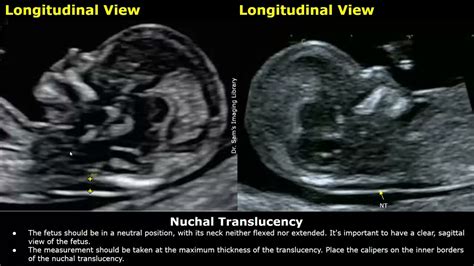
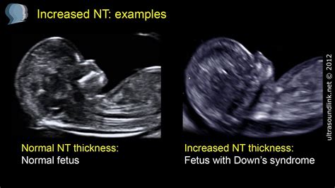
What is Nuchal Translucency (NT)? NT is the name given to the black area seen by ultrasound at the back of the fetal head/neck between 11 - 14 weeks of gestation. . There is a screening test, called the combined test, that determines this risk calculation. The test combines the measurement of the NT, the length of the baby, your age, and the .
Nuchal Translucency Normal Range Chart. When the nuchal scan is done, the doctor will share the results with you. At that time, it is important to understand what a normal measurement is. For a baby that is between 45 mm and 84 mm in size, a normal measurement is anything less than 3.5 mm. The NT grows in proportion to the baby.The nuchal translucency test measures the nuchal fold thickness. This is an area of tissue at the back of an unborn baby's neck. Measuring this thickness helps assess the risk for Down syndrome and otherThis is an area of tissue at the back of an unborn baby's neck. Measuring this thickness helps assess the risk for Down syndrome and other genetic problems in the baby. Alternative Names. Nuchal translucency screening; NT; Nuchal fold test; Nuchal fold scan; Prenatal genetic screening; Down syndrome - nuchal translucency. How the Test is PerformedThe nuchal translucency will examine: The progress of your pregnancy; The fluid underneath the skin along the back of the fetus’s neck, called the nuchal translucency, or nuchal fold. In some pregnancies, when the fetus has Down .
In another large series, the incidence of aneuploidy by nuchal thickness measurement was 7% with an NT between the 95th percentile for crown-rump length and 3.4 mm, 20% with NT between 3.5 mm and 4.4 mm, 50% with NT between 4.5 mm and 6.4 mm, and 75% with NT 8.5 mm or greater.Clinical resource with information about Thickened nuchal skin fold and its clinical features, . A thickening of the skin thickness in the posterior aspect of the fetal neck. A nuchal fold (NF) measurement is obtained in a transverse section of the fetal head at the level of the cavum septum pellucidum and thalami, angled posteriorly to .
Nuchal fold thickness is measured on an axial section through the head at the level of the thalami, cavum septi pellucidi, and cerebellar hemispheres (i.e. in the same plane that is used to assess the posterior fossa structures). One caliper should be placed on the outer edge of the skin, and the other against the outer edge of the occipital bone. The variables affecting nuchal skin fold thickness in mid trimester. Prenatal diagnosis. 2003; 23 (1):60–4. [Google . Bergman G, Almström H, Grunewald C, Valentin L. Is measurement of nuchal translucency thickness a useful screening tool for heart defects? A study of 16 383 fetuses. Ultrasound in obstetrics & gynecology. 2006; 27 (6 .Objective: To establish normal values of fetal nuchal fold thick-ness at 14-16 weeks of gestation by transvaginal sonography. Methods: Transvaginal sonography was used to measure nuchal fold thickness in 182 normal pregnancies at 14-16 weeks of gestation. Nuchal fold thickness was measured as the distance from the outer skull bone to the outer skin surface in the . Cho J, Kim K, Lee Y, Toi A. Measurement of nuchal skin fold thickness in the second trimester: Influence of imaging angle and fetal presentation. Ultrasound Obstet. Gynecol. 2005; 25 :253–257. doi: 10.1002/uog.1847.
In the 19 to 24 week gestational time frame, ≥6 mm appeared to be the optimal threshold, yielding a positive screen rate of 3.7% with a sensitivity of 83% (5/6). The adjusted positive predictive value was 1 in 38. The sensitivity of nuchal skin-fold thickness for Down syndrome detection was similar in women <35 and ≥35 years old.
Nuchal fold thickness increased with GA in a linear manner from 3.13 ± 0.68 mm (mean ± SD) at 16 weeks to 5.08 ± 0.76 mm at 24 weeks. The 95th percentile measurement at 24 weeks remained less than 6 mm. Conclusions. A threshold of 6 mm appears to be appropriate for the diagnosis of a thick nuchal fold even for gestations between 20 and 24 weeks.Sonographically measured nuchal skinfold thickness as a screening tool for Down syndrome: results of a prospective clinical trial. Obstet Gynecol. 1991; 77:533-536. . Sonographic measurement of nuchal skinfold thickness for detection of Down syndrome in the second trimester fetus: a multicenter prospective study. Obstet Gynecol. 1995; 85:103-106.
Nuchal translucency (NT) and nuchal fold thickening are two distinct soft markers for fetal abnormalities and aneuploidy.. The nuchal translucency is the normal fluid-filled space located at the back of the baby’s neck, and it is scanned via ultrasound during the first trimester. An increased nuchal translucency is considered a marker for fetal anomalies and can be .

thick nuchal fold normal baby
Resultado da iPad. A Espaçolaser, maior rede de depilação a laser do mundo, traz para você conveniência para agendar seus tratamentos e acompanhar suas sessões. .
nuchal skinfold thickness measurement|nuchal fold measurement 20 weeks