optic nerve thickness measurement mri|optic nerve size chart : distributing Explanation of how to measure and interpret optic nerve sheath diameter on . Sérgio Sacani Sancevero (Londrina, 17 de setembro de 1975), é um geofísico, youtuber, podcaster e proeminente divulgador científico brasileiro, dono do blog Space Today e apresentador do canal Ciência Sem Fim, associado aos Estúdios Flow.. Ver mais
{plog:ftitle_list}
webSaiba como pagar, recolher e abater o ISS da Prefeitura de Campina Grande, um tributo que incide sobre a prestação de serviços. Veja como gerar a NFS-e com ISS da .
Measurements of the optic nerve sheath diameter (ONSD) are most often taken at a distance of 3 mm from the posterior globe margin as this is believed to be the site of maximum pressure changes along the long axis of the optic nerve 1,2,4.
Explanation of how to measure and interpret optic nerve sheath diameter on .The optic nerve is the second (CN II) cranial nerve (TA: nervus opticus or nervus . We determined a threshold optic nerve area on MR imaging that predicts a clinical diagnosis of optic nerve atrophy and assessed the relationship between optic nerve area and retinal nerve fiber layer thickness measured . The optic nerve sheath diameter (ONSD) is an indirect marker of the intracranial pressure, but the normal range of ONSD as measured using magnetic resonance imaging .
The variations in the diameter of the optic nerve (ON) are important clinically in the diagnosis of conditions associated with the ON such as raised intracranial pressure, .
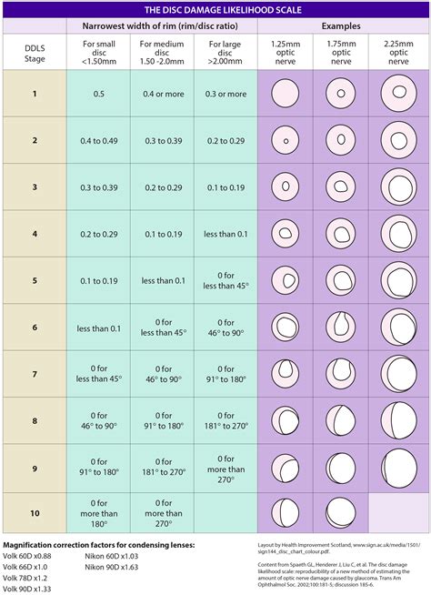
optic nerve size chart
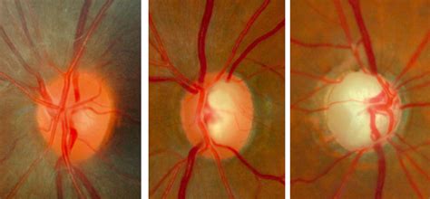
optic nerve different sizes
Optic nerve sheath diameter (ONSD) measurement may be a promising method for the non-invasive assessment of intracranial pressure (ICP) [1,2]. The sheath that surrounds .Explanation of how to measure and interpret optic nerve sheath diameter on ultrasound, MRI, and CT for assessing intracranial pressure. CONCLUSIONS: We report normative values of optic nerve diameter measured on MR imaging in children from birth to 18 years of age. A rapid increase in optic nerve diameter was demonstrated during the first 2 .Emerging MRI technologies that emphasize rapid acquisition should improve visualization of the optic nerve and facilitate accurate quantification of MRI properties that can detect visually-occult pathology.
Its main pathological changes are due to damage at the optic nerve head leading to loss of thickness of the RNFL. RNFL thickness can be measured using optical coherence .Optic Nerve Measurement on MRI in the S.R. Saaybi, A.M. Khamis and R.G. Hourani C.E. Al-Haddad, M.G. Sebaaly, R.N. Tutunji, C.J. Mehanna, . The aim of this study was to establish a data base for optic nerve diameter measurements on MR imaging in the pediatric population. . section thickness/spacing range 2–0.3 to 2–0.5 mm, acquisi- Table 1 shows the optic nerve mea-surements on MR imaging at the retro-bulbar and midaspect levels for each of the axial and coronal cuts. Optic nerve diameter measurements did not differ between the right and left eyes (Fig 2A) or between males and females (Fig 2B), regardless of the section and location of the measurement. However, the optic

BACKGROUND AND PURPOSE: No MR imaging measurement criteria are available for the diagnosis of optic nerve atrophy. We determined a threshold optic nerve area on MR imaging that predicts a clinical diagnosis of .MRI of the retrobulbar optic nerve was performed 5, 10, and 15 mm behind the eye with a 3-T whole body scanner (Magnetom Trio; Siemens, Munich, Germany). . Echographic measurement of optic nerve thickness correlated with neuroretinal rim area and visual field defect in glaucoma. Am J Ophthalmol. 1996;122:514–519. 21. KashiwagiK, OkuboT .
normal size of optic nerve
Background and purpose: No MR imaging measurement criteria are available for the diagnosis of optic nerve atrophy. We determined a threshold optic nerve area on MR imaging that predicts a clinical diagnosis of optic nerve atrophy and assessed the relationship between optic nerve area and retinal nerve fiber layer thickness measured by optical coherence tomography, an . Viewing the optic nerve through lenses and a slit lamp is the best way for your doctor to assess the optic nerve for glaucoma. Your doctor may document this assessment either with drawings or with optic disc photos. A color photograph provides a more accurate baseline for future comparison.
Optic nerve measurements were recorded 1 mm posterior to the optic disk. B. Prechiasmatic optic nerve measurements were recorded 5 mm posterior to the optic canal. C.Participant selection flowchart. OCT, optical coherence tomography. 2.2. SVD quantification. SVD quantification for the PREVENT Dementia cohort has been described elsewhere. 16, 17 Briefly, ePVSs were assessed separately in the basal ganglia and centrum semiovale in T2w MRI using a validated rating scale. 19 Scores ranged from 0 to 4 based on the number of lesions: 0 (none), . Epidemiology. In a study of 11 million people in the UK, optic neuritis (all causes) was found to have an incidence of 3.7 per 100 000 person-years. 69% of the cohort were female, with an average age of 35 years, and 92% were "white" 8. As typical optic neuritis is seen in the setting of multiple sclerosis (MS), most patients tend to be young adults, with a predominance .Purpose: To assess a novel magnetic resonance imaging (MRI) protocol for quantifying the optic nerve diameter (OND) as a measure of axonal loss in the optic nerve. Methods: Included in the study was one eye each from 47 subjects, of whom 9 had no eye disease, 16 had preperimetric glaucoma, 11 had a glaucomatous mean visual field defect of <10 dB and 11 of >10 dB.
instron tensile compression tester model 4481
ONSD MRI measurements of the transverse and sagittal diameters at a distance of 3 mm behind the papilla were evaluated twice each by two expert neuroradiologists. The correlations between MRI examiners were calculated using the concordance correlation coefficient (CCC). . Optic nerve sheath thickness is a promising non-invasive measure of . Eye angle exam. Corneal thickness measurement. Dilated eye exam. Eye pressure check.; Optic nerve imaging. Visual field test. What is glaucoma? Glaucoma is a term that includes several types of eye disorders that cause damage to your optic nerve.The condition is usually caused by increased pressure in your eye. Optic Nerve Atrophy Measurements with MRI in Patients with Diagnostic Utility of Optic Nerve Y.-M. Chang and R.A. Bhadelia B. Zhao, N. Torun, M. Elsayed, A.-D. Cheng, A. Brook, . equivocally consistent with optic nerve atrophy, and 2) RNFL thickness 85 m. Among these 52 patient eyes, a total of 34al MRI, especially since adjacent air-filled bony structures distort the MRI signal and motion is a problem even in cooperative, healthy volunteers. Evidence Acquisition: Over the past 3 years we have experimented with multiple novel MRI approaches and sequences to better characterize the optic nerves. The perfect MRI protocol would be quantitative and sensitive to subtle optic .
Its main pathological changes are due to damage at the optic nerve head leading to loss of thickness of the RNFL. RNFL thickness can be measured using optical coherence tomography (OCT), while the degeneration of the whole optic pathway can be assessed using MRI. . Diagnosis utility of optic nerve measurements with MRI in patients with optic . Optic nerve diameter on MRI correlates with Lumbar puncture opening pressure. . Normal measurement of optic nerve diameter ranges among literatures between values of 3–5.7 mm on . ROC curve analysis for neck fat thickness showed area under curve of 0.82 with the most accurate cut-off value at 1.1 cm at which the sensitivity is about 70 % .Measurement of optic nerve and optic nerve sheath was done on the 102 . slice thickness 2 mm, pixel bandwidth 150 Hz/pixel, FOV read 160 mm, . forward gaze and gentle eye closure during the scans. Axial scans were obtained parallel to the optic nerve. On MRI, the optic nerve sheath appeared as a high signal intensity surrounding the optic .
BACKGROUND AND PURPOSE: No MR imaging measurement criteria are available for the diagnosis of optic nerve atrophy. We determined a threshold optic nerve area on MR imaging that predicts a clinical diagnosis of optic nerve atrophy and assessed the relationship between optic nerve area and retinal nerve fiber layer thickness measured by . Purpose To determine a potential threshold optic nerve diameter (OND) that could reliably differentiate healthy nerves from those affected by optic atrophy (OA) and to determine correlations of OND in OA with retinal nerve fiber layer (RNFL) thickness, visual acuity (VA), and visual field mean deviation (VFMD). Methods This was a retrospective case control study. .
Abstract. Optic nerves are the second pair of cranial nerves and are unique as they represent an extension of the central nervous system. Apart from clinical and ophthalmoscopic evaluation, imaging, especially magnetic resonance imaging (MRI), plays an important role in the complete evaluation of optic nerve and the entire visual pathway. Normal appearance on MRI. Before being able to interpret MRIs of the region it is important to understand the normal anatomy of the pituitary gland and surrounding structures: pituitary gland. cavernous sinus. optic nerve/optic chiasm/optic tract. suprasellar cistern. third ventricle. The anterior and posterior parts of the pituitary gland are .Radiopaedia.org, the peer-reviewed collaborative radiology resource The peripapillary retinal nerve fibre (pRNFL) thickness is a measure that can be used to quantify axonal integrity in pathological processes involving the optic nerve. . and optic nerve glioma .

Nerve Fiber Analyzer (GDx): GDx uses laser light to measure the thickness of the nerve fiber layer. There are nuances between the instruments, but they all serve the purpose to quantitatively analyze the nerve fiber layer thickness and measure certain parameters of the optic nerve head and macula. To examine the relationships of retinal structural (optical coherence tomography) and visual functional (multifocal visual evoked potentials, mfVEP) indices with neuropsychological and brain structural measurements in healthy older subjects. 95 participants (mean (SD) age 68.1 (9.0)) years were recruited in the Optic Nerve Decline and Cognitive Change (ONDCC) .o Table of measurements: RNFL thickness and RNFL symmetry* ONH analysis: o Table of measurements: optic nerve rim area, disc area, average and vertical C:D, and cup volume. o Neuro-retinal rim thickness plots* Dimensions of analysis area—centered on fovea: 2 mm x 2.4 mm. Macula analysis: o GC-IPL thickness maps. o Horizontal B-scans
normal optic nerve thickness
Nerve protrusion length measurement by MRI is based on a previously described MRI measurement of globe deformation that converts the 3D geometry of the globe's posterior wall into a 2D map of the distances from the center of the globe to the head of the optic nerve and the surrounding region. 20 The MRI-based measurement of NPL (NPL-MRI) was .
A comitiva volta para a fazenda e José Leôncio não encontr.
optic nerve thickness measurement mri|optic nerve size chart